Why caries is easy to miss - even for experienced dentists
Research shows dentists miss more than half of initial caries lesions on radiographs - and that detection drops sharply under time pressure. A look at why the task is genuinely hard, and what can realistically help.
Radiographs are commonly used to help detect and diagnose caries, especially the approximal lesions between teeth, which are often not detectable by visual-tactile examination alone.1–4 However, research shows that dentists reading X-rays miss many lesions. Here we aim to shed light on what may lead to those missed pathologies and what could be done to improve dentists' and patients' experience and outcomes.
Missed findings are common, even in ideal conditions
The data shows that more than half of initial4–6 and nearly half of the secondary7 lesions are missed on radiographs.
Whether a lesion is detected also depends on who reads the radiograph, and when, because agreement between readers and within the same reader over time is limited.8 Experience helps, but it does not remove the problem - dental students, dentists, and radiologists all miss lesions.9 It is more difficult to find early enamel lesions on periapical radiographs than on bitewings, indicating that how well a lesion shows up on the image also matters, not only who reads it.10
These misses happen in research settings, without the stress of having patients waiting, which is commonplace for practising dentists. This suggests that the task is hard, not that dentists lack skill.
Time pressure makes it harder
Tight schedules and stress complicate it even further. A 2019 survey11 found high levels of stress and burnout, with general dental practitioners most affected. Yet until recently, almost no experimental research had looked at how stress affects clinical performance.12
In 2019 it was tested directly.13 Dentists reported on two sets of bitewings, one set with a countdown clock and one without.
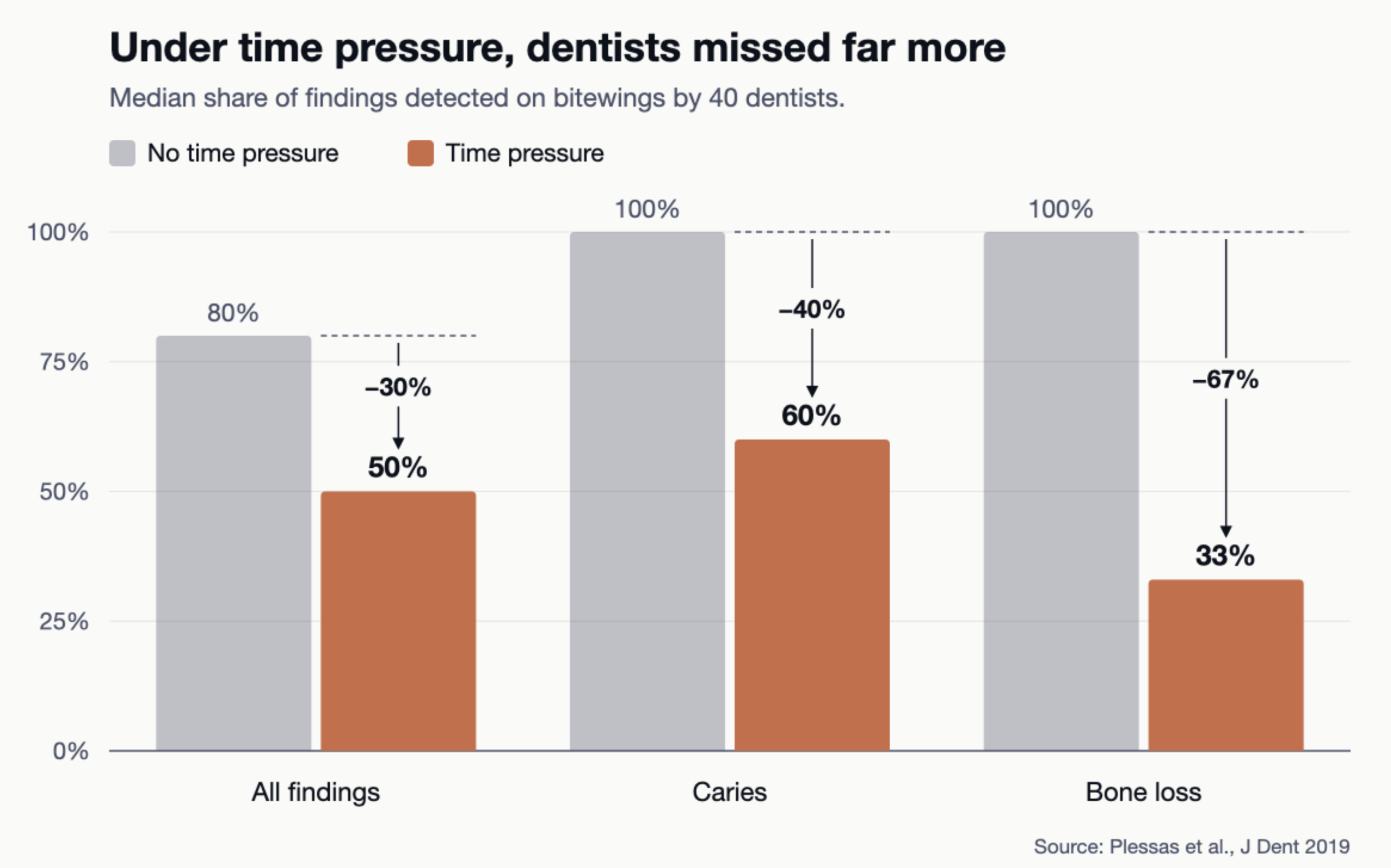
Under time pressure, they missed significantly more pathologies, while they hardly ever reported pathologies that weren't there. The authors read this as dentists missing information rather than misreading it.
What dentists tell us
In our conversations with dentists, the same three points come up again and again.
- Light on the screen. Light in the room makes the screen harder to read.
- Time constraints. There is little time to look at each image.
- Back-to-back patients. One patient follows the next, with no gap to go back over an image.
Screens near a window or under more ceiling lights can show fewer faint details on a test image, and screens can vary widely,14 which may lead to missed early enamel lesions. Another study showed that room light significantly changed how confident radiologists felt, and referred to earlier studies suggesting that brighter rooms made caries harder to detect.15
Time pressure has a significant effect on what dentists miss.13 A packed schedule affects patients too. Waiting times and the time and attention dentists give during appointments shape patient satisfaction and trust, which in turn are linked to patients returning for care.16 Time per patient is therefore not only a question of what the dentist sees on the radiograph, but of how patients experience their care.
What can help
The study's authors suggest two practical steps: longer appointments when radiographs are taken, and reviewing radiographs again later, away from time pressure. Both cost time, which is the resource that is already scarce in busy practice.13
Decision support software is another option. When dentists read bitewings with and without AI support, the share of caries lesions detected rose significantly from 72% to 81% with AI, without more false alarms.17 The higher detection rate is valuable, but dentists using AI also made more invasive treatment decisions, which shows that clinical assessment remains central. AI should support the dentist's judgement, not replace it.
Missed findings on radiographs are a shared, well-documented problem, rooted in how hard the task is and the conditions dentists work in. AI decision support offers a second reader that is not affected by fatigue,4 a full schedule, or a poorly lit treatment room. Used to support the dentist's judgement, not replace it, AI can help make missed findings less likely.17
Sources
- Köse E, Hoşgör S. Assessment of posterior interproximal surfaces on bitewing and panoramic radiographs by dental interns and an artificial intelligence application. Oral Radiol. 2026;42(2):400-412. doi:10.1007/s11282-025-00872-9
- Wang Y, Fu D, Zhou G, Zou L. AI-based detection of proximal caries across radiographic modalities: a systematic review and meta-analysis. Dentomaxillofacial Radiol. Published online July 6, 2026:twag046. doi:10.1093/dmfr/twag046
- Heiden G, Pereira P, Yoshida ML, Oliveira D, Rocha M, Ribeiro APD. Comparative evaluation of proximal caries detection methods using human, artificial intelligence, and micro-CT assessments. J Dent. 2026;173:106835. doi:10.1016/j.jdent.2026.106835
- Ganss C, Jung K, Schilling L, Sonderegger S, Neuhaus KW. Human and artificial intelligence performance in radiographic caries detection: ex vivo tooth section-referenced evaluation and implications for clinical decision-making. J Dent. 2026;168:106602. doi:10.1016/j.jdent.2026.106602
- Walsh T, Macey R, Riley P, et al. Imaging modalities to inform the detection and diagnosis of early caries. Cochrane Database Syst Rev. 2021;2021(3):CD014545. doi:10.1002/14651858.CD014545
- Cantu AG, Gehrung S, Krois J, et al. Detecting caries lesions of different radiographic extension on bitewings using deep learning. J Dent. 2020;100:103425. doi:10.1016/j.jdent.2020.103425
- Brouwer F, Askar H, Paris S, Schwendicke F. Detecting Secondary Caries Lesions: A Systematic Review and Meta-analysis. J Dent Res. 2016;95(2):143-151. doi:10.1177/0022034515611041
- Natto ZS, Olwi A, Abduljawad F. A comparison of the horizontal and vertical bitewing images in detecting approximal caries and interdental bone loss in posterior teeth: A diagnostic accuracy randomized cross over clinical trial. J Dent Sci. 2023;18(2):645-651. doi:10.1016/j.jds.2022.08.006
- Gonzalez-Valenzuela RE, Vu QTD, Mettes P, Loos BG, Marquering H, Berkhout E. Human-AI Collaboration in Proximal Early Caries Lesion Detection: Potential Benefits for Novices and Potential Risks for Experts. Caries Res. Published online September 10, 2026. doi:10.1159/cre/abuag009
- Takahashi N, Lee C, Da Silva JD, et al. A comparison of diagnosis of early stage interproximal caries with bitewing radiographs and periapical images using consensus reference. Dentomaxillofacial Radiol. 2019;48(2):20170450. doi:10.1259/dmfr.20170450
- Collin V, Toon M, O'Selmo E, Reynolds L, Whitehead P. A survey of stress, burnout and well-being in UK dentists. Br Dent J. 2019;226(1):40-49. doi:10.1038/sj.bdj.2019.6
- Plessas A, Delgado MB, Nasser M, Hanoch Y, Moles DR. Impact of stress on dentists' clinical performance. A systematic review. Community Dent Health. 2018;35(01):9-15. doi:10.1922/CDH_4118Plessas07
- Plessas A, Nasser M, Hanoch Y, O'Brien T, Bernardes Delgado M, Moles D. Impact of time pressure on dentists' diagnostic performance. J Dent. 2019;82:38-44. doi:10.1016/j.jdent.2019.01.011
- Hastie T, Venske-Parker S, Aps JKM. Impact of viewing conditions on the performance assessment of different computer monitors used for dental diagnostics. Imaging Sci Dent. 2021;51(2):137-148. doi:10.5624/isd.20200182
- Liu XX, Li G. Diagnostic performance of approximal caries in bitewing radiographs from different monitors and room illuminances. Dentomaxillofacial Radiol. 2025;54(2):125-131. doi:10.1093/dmfr/twae061
- Haji Seyed Javadi SK. From Patient Experience to Dental Service Return Visits: A Narrative Review of Strategies for Improving Dental Care Services. J Patient Exp. 2025;12:23743735251406349. doi:10.1177/23743735251406349
- Mertens S, Krois J, Cantu AG, Arsiwala LT, Schwendicke F. Artificial intelligence for caries detection: Randomized trial. J Dent. 2021;115:103849. doi:10.1016/j.jdent.2021.103849